
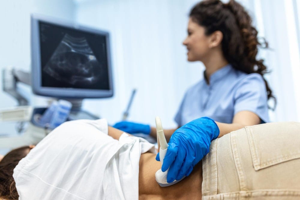
Examination by ultrasound
Ultrasound
Ultrasound examination is one of the safest and most accessible diagnostic methods in modern medicine. It does not burden the body with radiation, it is painless and you know...
Regular breast examination remains one of the most important steps in the prevention of breast cancer. Two main methods are most often compared - breast ultrasound and mammography. Each has its strengths and ideal situations in which it excels. At Hello Beauty Clinic, we offer high-quality breast ultrasound on a modern, high-resolution device.
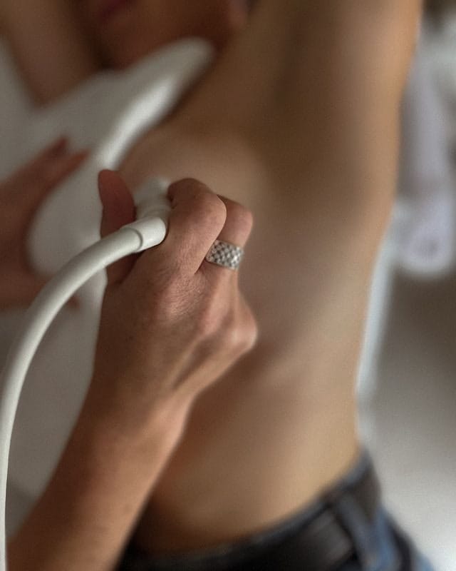
Breast ultrasound works with ultrasound waves that pass through tissue, reflect and create a detailed image in real time. The examination is performed lying down, a conductive gel is applied to the skin and the doctor moves the probe over the breast and armpit. It usually takes 15-20 minutes, is painless and does not require any compression of the breast.
Main advantages of breast ultrasound
Thanks to these properties, ultrasound is considered a very accurate and gentle method - especially for younger women and women with dense breast tissue. In dense tissue, which often makes it difficult to detect lesions on mammography, ultrasound penetrates much better and detects changes with higher sensitivity.
The examination is comfortable – no compression, no radiation, no preparation. The doctor can assess the mobility of the lesion, blood supply or tissue stiffness in real time, which provides additional important information. At Hello Beauty Clinic, we use only the most modern high-resolution devices for maximum detail.
A mammogram is an X-ray examination of the breasts. The breast is gently pressed between two plates for a short time to minimize blurring, and images are taken from several angles. Modern digital machines use a low dose of radiation and provide significantly more detailed images than older analog systems.
Main advantages of mammography
In addition, modern 3D tomosynthesis increases the accuracy of detection, reduces the number of false positives and better handles even slightly denser tissue compared to classic 2D mammography. Nevertheless, in women with significantly dense glandular tissue (often up to 45–50 years old), it is usually supplemented with ultrasound to achieve the most accurate result.
| Parameter | Breast Ultrasound | Mammography |
|---|---|---|
| Principle | Ultrasound waves | X-ray radiation |
| Radiation | None | Low dose |
| Best for | Dense / glandular tissue, younger women | Fatty tissue, women over 50 years |
| Detection of microcalcifications | Weaker | Excellent |
| Painfulness | None | Mild (breast compression) |
| Examination time | 15–20 minutes | 10–15 minutes |
| Immediate result | Yes | No (report usually within a few days) |
| Screening role | Supplementary, preventive for younger women | Primary screening from age 45 |
A breast ultrasound is in many cases the method of first choice, especially if you are younger than 45–50 years old. At this age, dense or moderately dense glandular tissue prevails in most women, which is best assessed by ultrasound - it can penetrate the layers of tissue much more accurately than mammography.
Ultrasound is also an ideal choice if you feel any discomfort - breast pain, swelling, a palpable mass, a change in shape or asymmetry of the breasts. In such situations, it allows you to quickly and reliably assess whether it is a harmless change or something that requires further examination.
Another important group are women who are pregnant, breastfeeding or for any reason prefer an examination completely without ionizing radiation. Ultrasound is completely safe in these cases and does not burden the body.
Many women also appreciate the speed and convenience - the examination takes only 15–20 minutes, is painless and immediately after it the doctor will explain the result to you and, if necessary, suggest further procedures. You don't have to wait for a description of the images for several days. Moreover, with us, no request form from a general practitioner is required - you just need to make an appointment and come for the examination.
It is precisely thanks to the high accuracy in dense tissue, the complete absence of radiation and the great comfort that many women consider ultrasound to be the method of first choice in these situations.
Ultrasound examination of the breast is also a very suitable method for women with breast implants. It can diagnose with high accuracy an implant rupture with silicone leakage, a change in the position of the implant, seroma (fluid around the implant) or hematoma (bleeding).
Yes – for dense tissue, which is common in women under 45–50 years of age, ultrasound has a higher sensitivity than mammography alone.
The dose is very low. The benefit of regular screening significantly outweighs the theoretical risk.
As a preventive measure, once a year for women under 45–50 years of age (or as recommended by a doctor). Immediately if there are any problems.
Ultrasound is very convenient and problem-free. Mammography can be performed, but it requires a special technique.
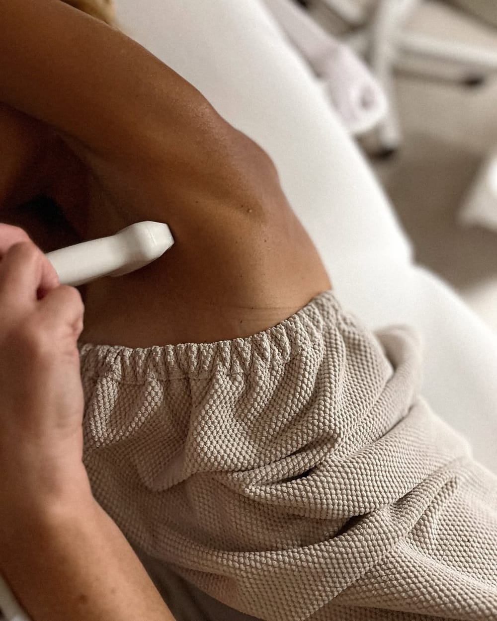
Each of the methods - breast ultrasound and mammography - has its place in prevention and diagnosis. Mammography remains the basic screening method from the age of 50 thanks to its proven effect on reducing mortality and the best detection of microcalcifications. Ultrasound, on the other hand, excels in women under 45–50 years of age and with dense glandular tissue, where it offers higher accuracy, zero radiation exposure and immediate results with the possibility of detailed assessment in real time.
There is no universally better method - your age, type of breast tissue and current needs are decisive. At Hello Beauty Clinic, we focus on high-quality breast ultrasound on a modern device, without the need for a request form and with enough time to explain the results.
Ultrasound examination is one of the safest and most accessible diagnostic methods in modern medicine. It does not burden the body with radiation, it is painless and you know...
Excessive sweating of the whole body is not only an aesthetic problem. It can make our daily life, both personal and social, uncomfortable. It also very often leads to...
Proper skin care consists of four basic steps: cleansing, moisturizing, sun protection, and regular exfoliation. These steps apply to every skin type, only the choice of...