Pancreas ultrasound
Pancreatic ultrasound (pancreatic sonography) is a modern, painless and accurate examination. It allows you to examine the pancreas without radiation exposure. At Hello Beauty Clinic, we offer this examination as part of preventive and diagnostic care. The entire process takes 15 to 30 minutes. You will know the result immediately. An ideal choice for those who want to be sure about the condition of the pancreas. In some cases, part of the pancreas may be inaccessible for ultrasound examination and it is necessary to examine it with another imaging method (CTR, MR).
Pancreas in the body and why you should check it regularly
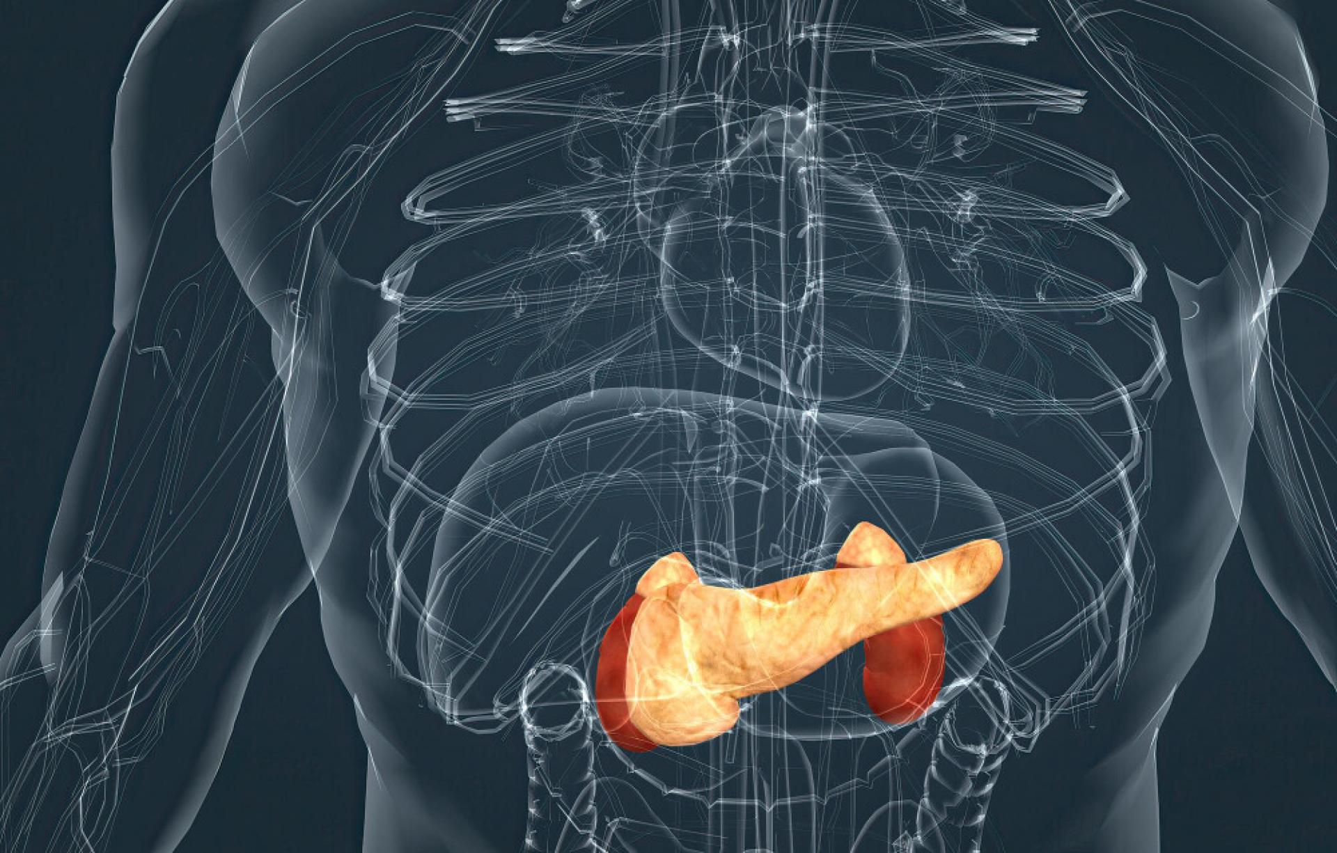
The pancreas is an organ located deep in the upper abdomen behind the stomach. It performs two essential functions that affect overall health.
The exocrine part of the pancreas produces digestive enzymes (amylase, lipase and proteases). These enzymes travel through the main duct to the duodenum, where they help break down carbohydrates, fats and proteins from food. Without sufficient enzymes, digestion disorders, bloating or inadequate absorption of nutrients occur.
The endocrine part consists of the islets of Langerhans. This is where key hormones are produced - insulin, which lowers blood sugar levels, and glucagon, which, on the contrary, increases them. A malfunction of this function leads to diabetes or other metabolic problems.
Pancreatic disorders often occur for a long time without significant symptoms. The most common include acute or chronic inflammation (pancreatitis), cyst or pseudocyst formation, benign and malignant tumors, and changes associated with long-term diabetes. The risk is increased by excessive alcohol consumption, smoking, high blood fat levels, or gallstones.
Many patients only experience symptoms during an acute attack – sudden sharp pain in the upper abdomen that radiates to the back, intense nausea, vomiting, fever, or unexplained weight loss. In advanced cases, serious complications such as tissue necrosis or the development of diabetes are at risk.
Regular pancreatic ultrasound allows these changes to be detected at an early stage, when they do not yet cause significant problems. Early detection is often sufficient to adjust lifestyle, treat, or monitor. This prevents acute attacks, long-term damage, and the need for more invasive examinations. Prevention is truly the key to maintaining quality of life.
What changes does an ultrasound of the pancreas most often reveal
Modern devices provide a clear picture of the size, shape, structure and surroundings of the pancreas. The doctor assesses several parameters during the examination.
Possible findings include:
- Acute or chronic inflammation (pancreatitis)
- Cysts and pseudocysts
- Thickening or irregularities in the structure
- Tumor foci
- Dilation of the pancreatic duct (ductus pancreaticus)
Thanks to high resolution, we can detect even minor abnormalities. At our clinic, we use the latest generation equipment for maximum accuracy.
Are you interested in a pancreas examination?
*Please book for morning appointments and arrive on an empty stomach.
When is it appropriate to have your pancreas checked
Pancreatic problems often only become apparent when they are already quite serious. The first signs include unexplained digestive problems - for example, a feeling of fullness after a small amount of food or sudden weight loss without changing your diet.
The pain is typically in the upper middle or left abdomen, may radiate to the back, and may worsen after eating or drinking alcohol. It is often felt as a dull ache rather than a sharp pain. Other warning signs include new-onset diabetes in adults over 40, recurrent nausea without an obvious cause, or skin changes (jaundice, itching).
We also recommend testing for people who have a history of heavy drinking, smoking, high blood fat levels, or a history of viral hepatitis. If blood tests show elevated pancreatic enzymes (amylase, lipase), an ultrasound is the first step to clarify the cause.
Preparing for the examination
A quality image of the pancreas requires minimal preparation. Come on an empty stomach – do not eat for at least 6-8 hours before the appointment. The day before, avoid carbonated drinks, legumes and heavy meals. Take your usual medications with a small amount of plain water. If you have specific restrictions, let us know when ordering. This will help us get the most accurate image possible.
How the pancreas examination is performed
The examination takes place in a quiet environment. You lie on a bed on your back or slightly on your side and the doctor applies a transparent gel to your upper abdomen.
The probe gently moves over your skin and sends ultrasound waves. The image is displayed in real time on a monitor. The doctor examines the pancreas from multiple angles and occasionally asks you to hold your breath or change position for better visibility.
The entire process is painless. You will only feel light pressure and the coolness of the gel. After the examination, the doctor will immediately explain the results and answer your questions. If necessary, we will suggest further steps or a consultation with a gastroenterologist.

Spleen sonography
At our clinic, we also offer spleen sonography as part of abdominal sonography. This organ is located in the upper left quadrant of the abdomen, right next to the pancreas and stomach. The spleen filters the blood, removes old red blood cells and participates in the immune response.
Spleen examination can be performed simultaneously with pancreatic sonography or as a separate examination. It allows to detect spleen enlargement (splenomegaly), cysts, masses, hematomas after injuries or changes in blood, liver or infection diseases (e.g. mononucleosis).
Typical symptoms that lead to spleen examination include a feeling of fullness or pressure in the left lower rib cage, pain when taking a deep breath, fatigue or a tendency to bruise and bleed easily. The risk of spleen enlargement increases with liver diseases (cirrhosis), blood disorders, lymphomas or after previous infections.
The preparation and course of the spleen examination are the same as for the pancreas – fasting or a light meal beforehand and a comfortable position on the bed. The doctor will move the probe slightly to the left and examine the spleen from multiple angles. The total time will be extended by only a few minutes.
If you suspect problems with the spleen or want a comprehensive view of the left upper quadrant of the abdomen, we will be happy to provide you with a combined examination of the pancreas and spleen. This will give you a comprehensive picture of the condition of both organs in one appointment.
Why choose a pancreatic ultrasound with us
The examination is safe for anyone, including pregnant women. We do not use ionizing radiation, so it can be repeated as needed.
Clients will appreciate quick appointments, immediate evaluation and a pleasant environment. Our specialists have many years of experience in abdominal sonography and an individual approach.
Pancreatic ultrasound is therefore a simple way to gain peace of mind and certainty about the health of this sensitive organ.
Pancreatic ultrasound is an investment in smooth digestion, stable sugar levels and prevention of serious complications. At our clinic, we combine cutting-edge technology with a human approach. A healthy pancreas means more energy, fewer problems and a sense of security every day.
Take a step towards better health – book a pancreatic ultrasound.
*Please book for morning appointments and arrive on an empty stomach.